
My work for the Phil and Penny Knight Campus for Accelerating Scientific Impact has allowed a number of professional explorations I had never considered or had opportunity to do.
For the past two years, I’ve had to adjust my senses and visualization skills to the idea of capturing research practice and science. And while many of the physical settings are the same, the challenge has been to find different ways to capture objects, settings, and people in ways that are revealing and feel fresh.
One particular challenge that has been fascinating to explore is how to help visualize the science behind the actual research. On some occasions, macro photography has been very helpful in capturing physical objects and small devices, but in most situations, especially for bioengineering, either the research occurs exclusively at a micro level or the chemistry behind the work is considerably abstract.
In order to build more research assets for the Knight Campus, I began pushing the limits of what I knew and could do with a camera. I began playing with microscopes and conceptual 3D modelling to help illustrate the science behind certain projects.
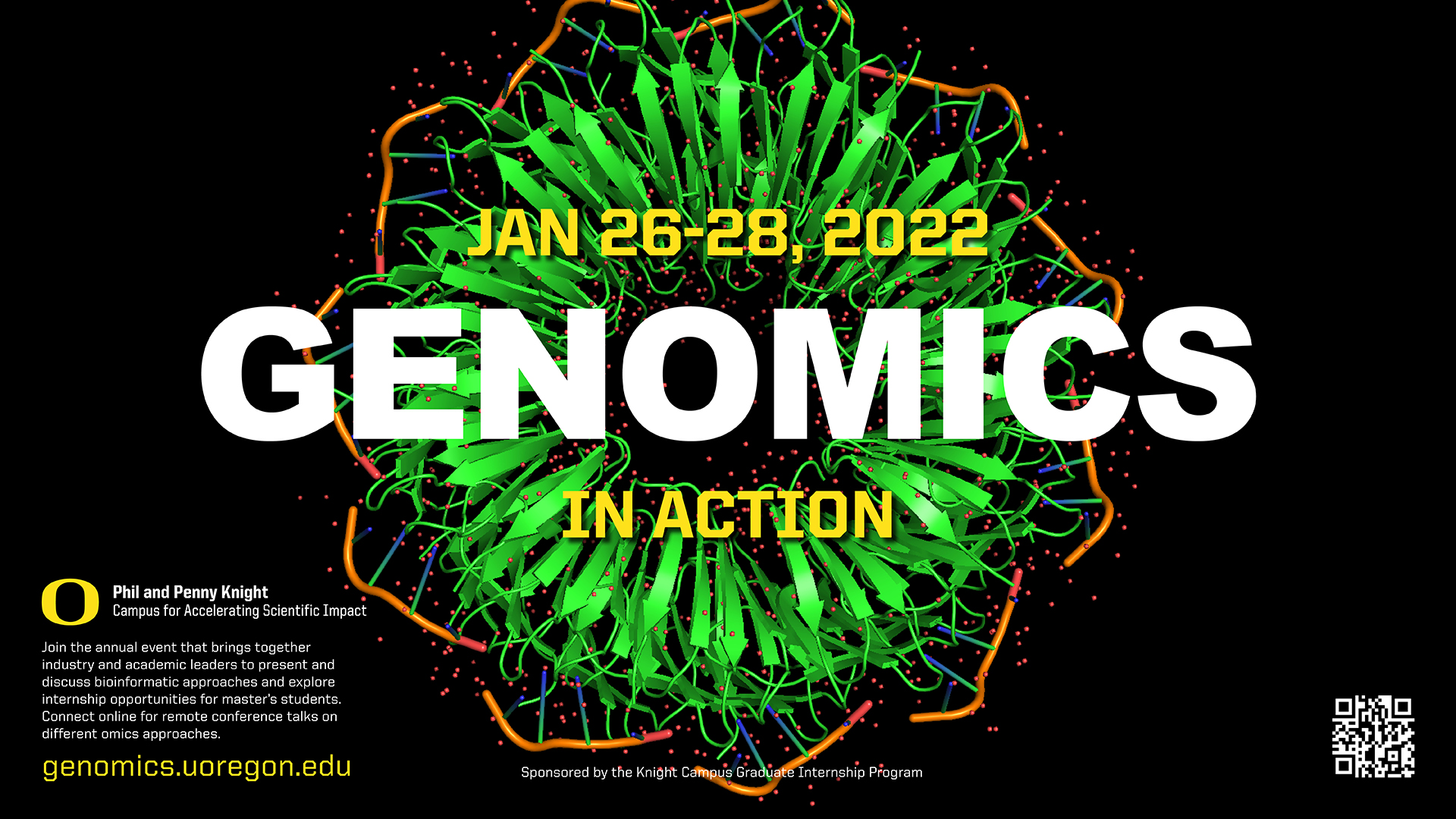
One of the first things I tried my hand at was learning the basics of how to open Protein Data Bank (PDB) files into Blender 3D and apply my modelling skills so I could help illustrate some science work or create collaterals using such images. The initial learning curve was a bit brutal, but I quickly understood ways to use the Research Collaboratory for Structural Bioinformatics PDB and find different types of proteins or molecules for inspiration. At first, the exercise was strictly learning how to model, color grade, and export renders of proteins. Later, I began using the concept in actual digital collaterals for promoting bioinformatics.

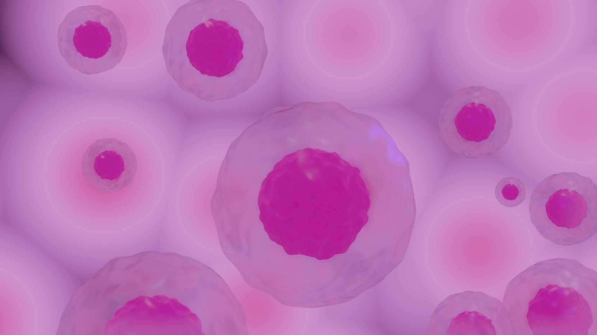

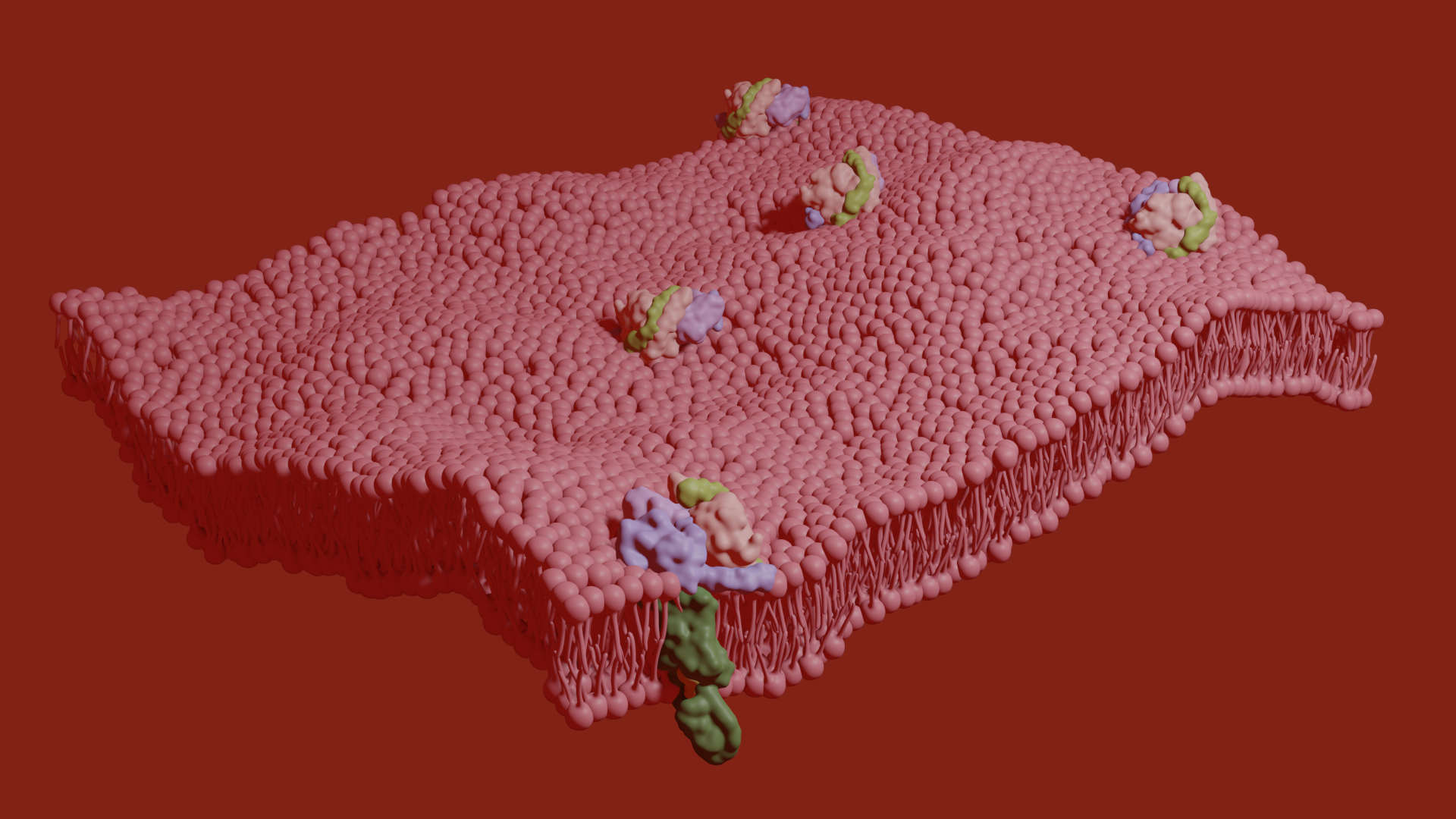
I also began exploring actual scientific visualization tools and image analysis programs like Avogadro, Chimera X, Fiji, and PyMol. These analysis programs are intended to help scientists visually analyze and demonstrate chemical reactions at a molecular level. For me, they became a treasure trove of visual information and aesthetic possibilities beyond what a traditional camera could help me capture or illustrate.
An interesting experience was gaining access to lab spaces and tools I had never seen, much less used. It has been amazing to collaborate with different lab managers in these settings and learn how to best use certain tools to capture the research being conducted. In my time at the Knight Campus (and despite the pandemic) I have learned to use a stereoscope, spent time inside a nanofabrication lab, and even learned best practices for how to build a PRUSA 3D printer.
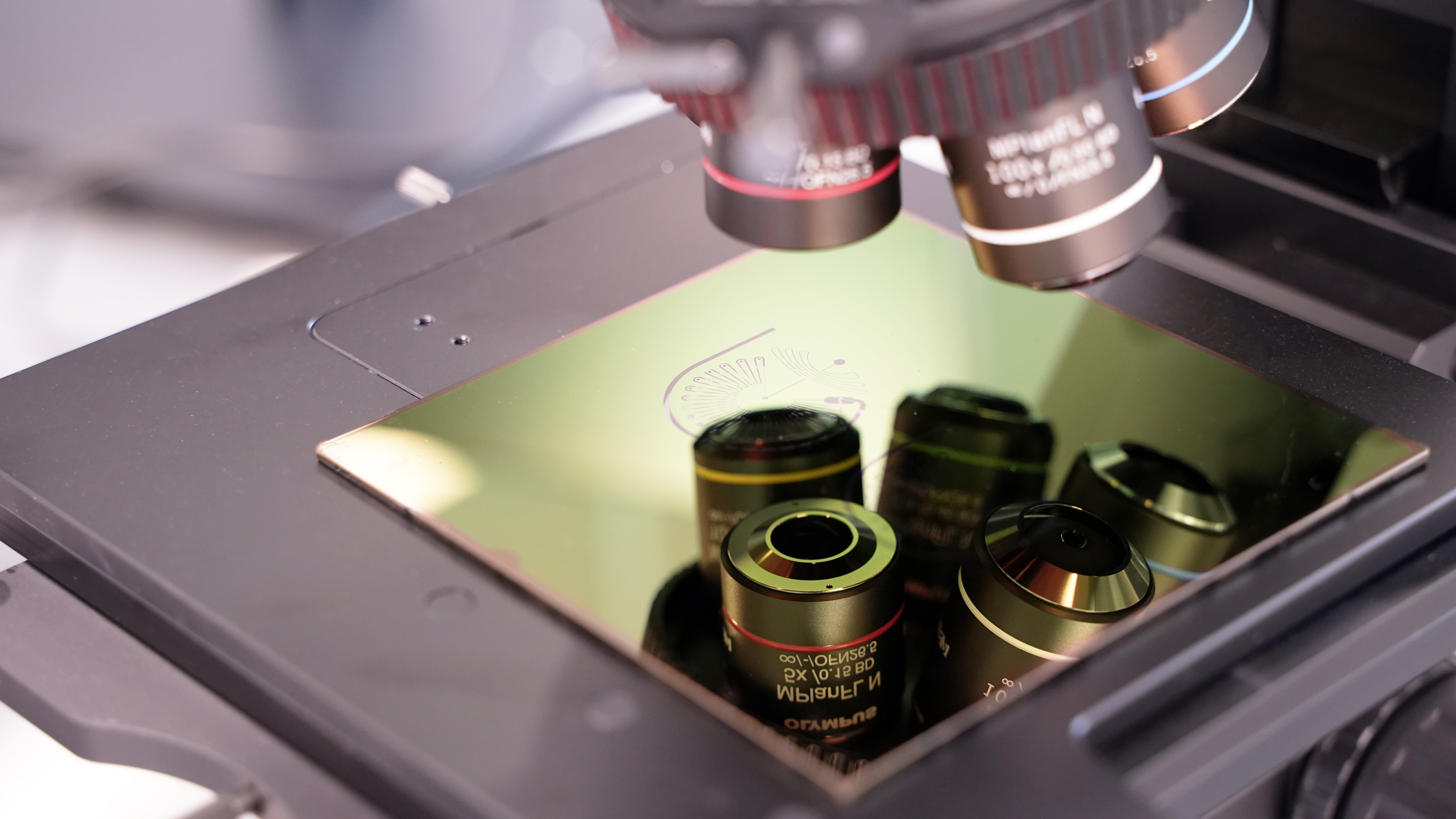

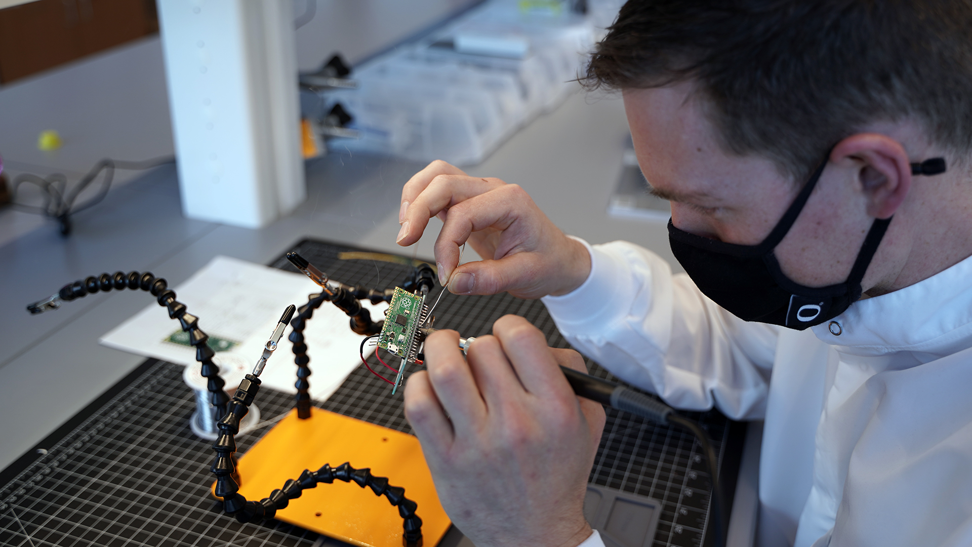
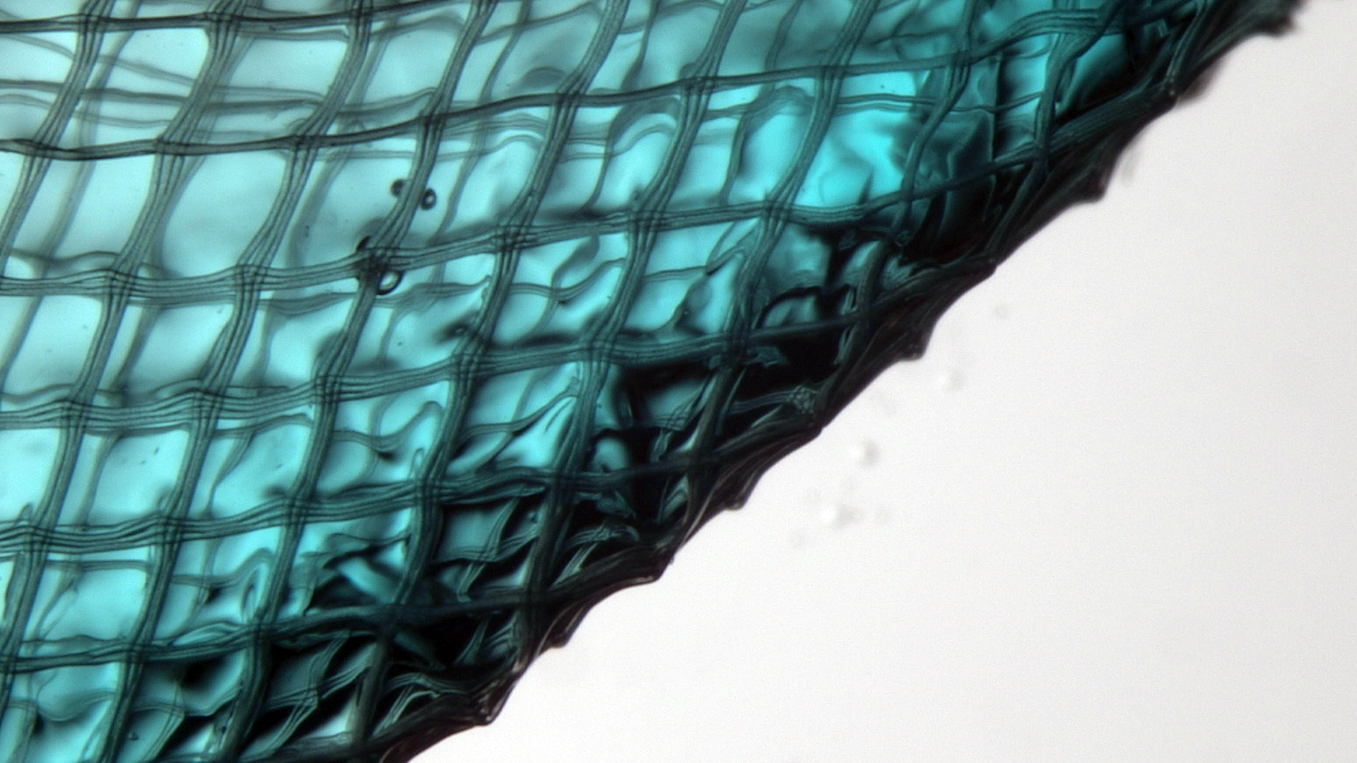
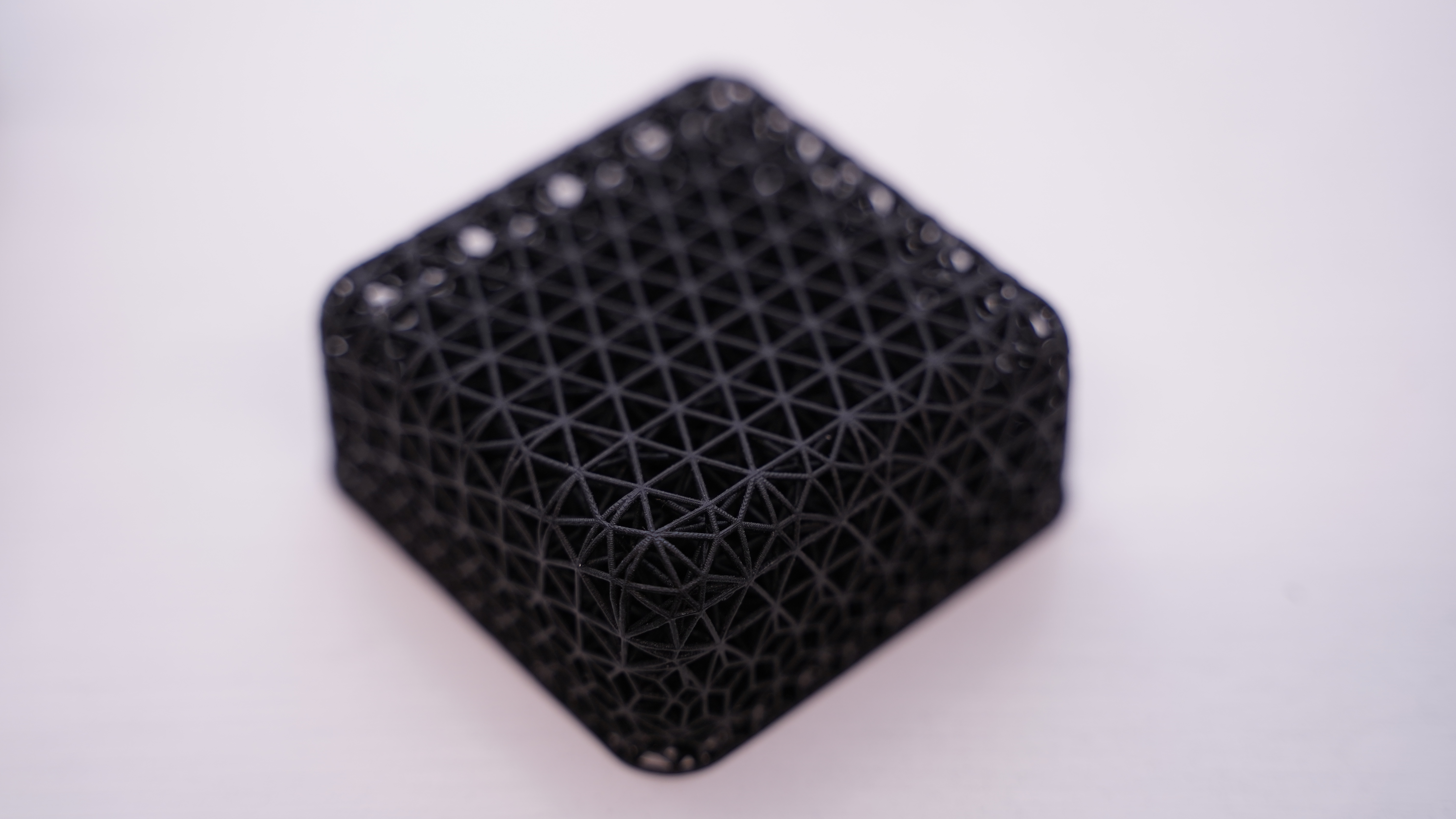
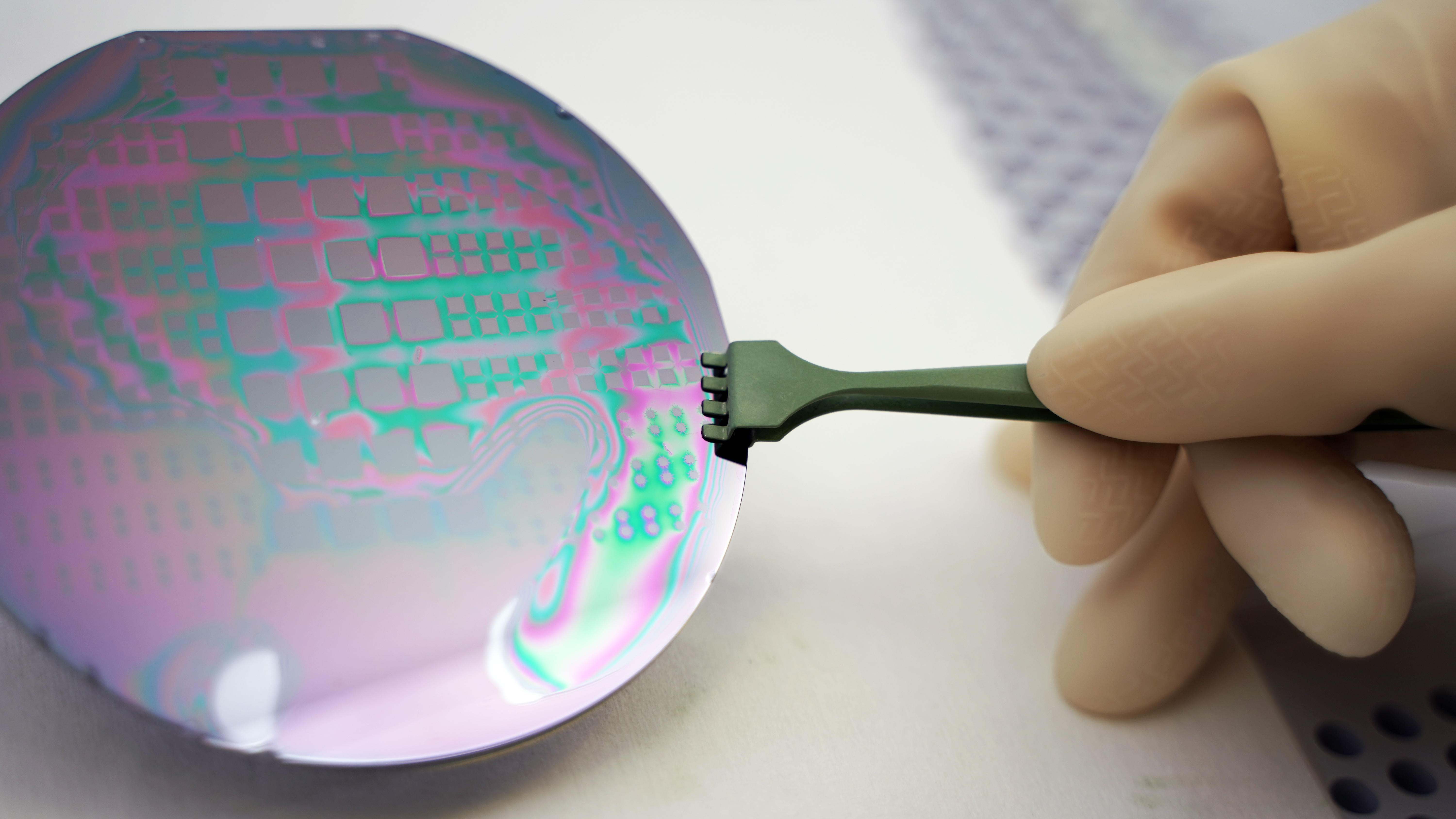
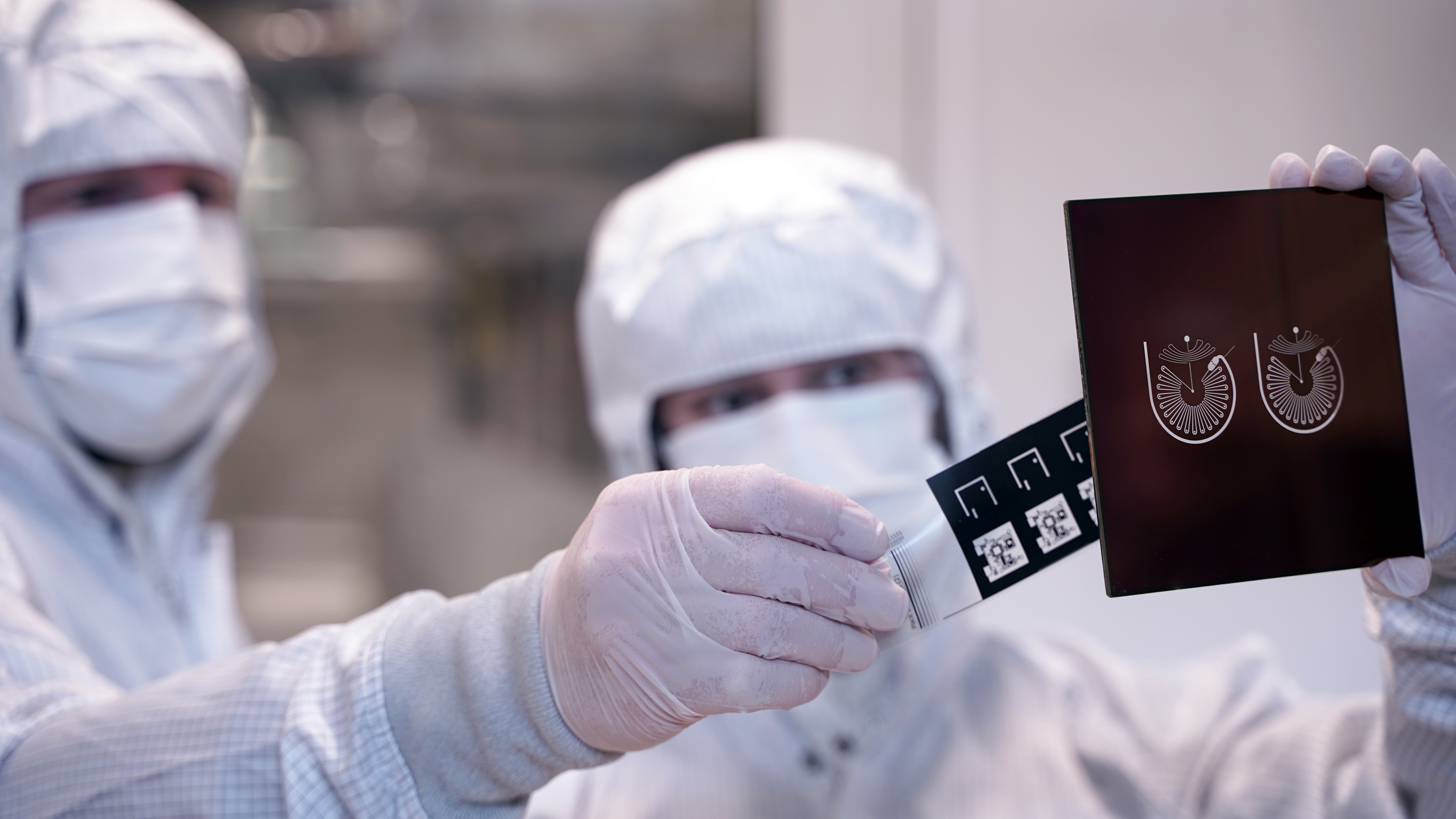
Photo below: Knight Campus students Yan Pacheco and Anissa Benabbas holding a set of lithography prints.
My process may have begun with a small amount of frustration, but in the end, it has allowed me to explore a number of things I had never accomplished and learn to use tools either unfamiliar to me, or way out of reach in regular design practice.
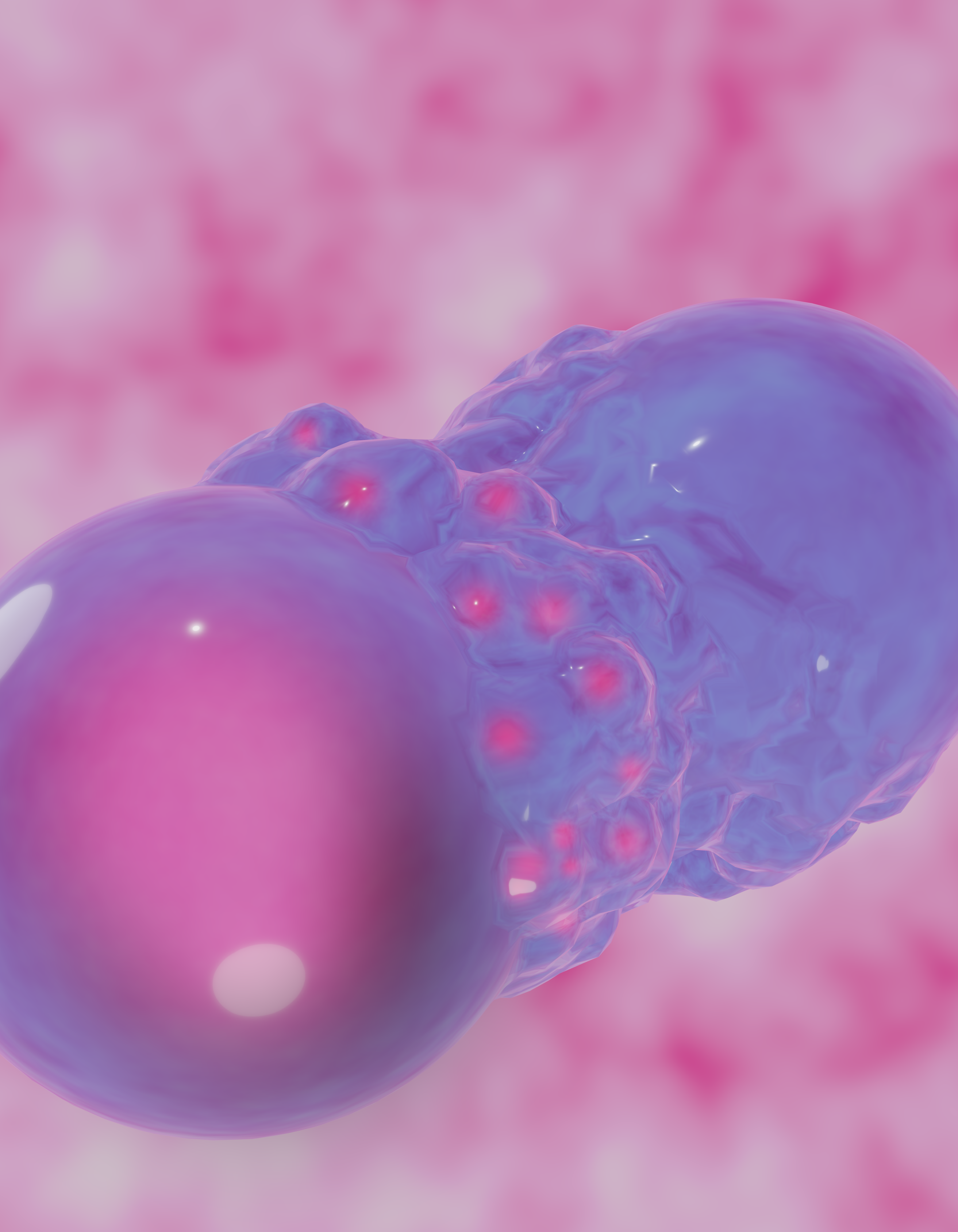
The high mark of my work on science photography and illustration is without a doubt my collaboration with Professor of Bioengineering at the Knight Campus, Dr. Gabriella Lindberg. She had secured publication of her latest paper through Advanced Materials Interfaces Journal and requested the opportunity to compete for a journal cover. We discussed several concepts and ended-up submitting our fourth concept. We got approved almost immediately. This was my first molecular biology illustration submission and it was published as a frontispiece on November 3, 2022 edition of Advanced Materials Interfaces.
While I do not call myself a true Science Illustrator, I feel confident I can hold my own using traditional photography skills, microscopic cameras, molecular analysis tools, and 3d modelling programs to help visualize different types of science research.
During the pandemic I had the opportunity to build a full desktop computer for my son Darian. I hope to have an excuse some time soon to build my own PRUSA 3D printer!
